MolNFT v1 · #183190 · protein
7NHT: AKIRIN2 bound human proteasome
7NHT · Proteasome subunit alpha type-2: “AKIRIN2 bound human proteasome”. Electron microscopy, 1 ligand (K), 20 pockets (best druggability 0.84). MolNFT v1 #183190 on GenesisL1: 3D structure, binding sites and pockets in GL1X.
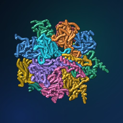
- PDB
- 7NHT
- Type
- protein
- Length
- 3,222 residues
- Chains
- 16
- Method
- electron microscopy
- Deposited
- 11-FEB-21
- Collection
- MolNFT v1 / PDB
- Contract
- 0xDE3723766Bc32dcACD03C17BaA400A7B36837Eba
- Owner
- 0x3396CDB397284571d12822B4ae1189a8E22AAAAA
- Molecule
- Proteasome subunit alpha type-2; Proteasome subunit alpha type-4; Proteasome subunit…
- EC
- 3.4.25.1
- UniProt
- P25787, P25789, O14818, P28066, P25786, P25788, P60900, Q99436, P49720, P49721, P28074, P20618, P28070, P28072, Q53H80
- Ligands
- K
- Pockets
- 20 · best druggability 0.84
Structure
electron microscopy · 16 distinct chains · 36% helix, 28% strand (DSSP).
| Chain | Molecule | Type | Residues | Secondary structure | UniProt |
|---|---|---|---|---|---|
| A | Proteasome subunit alpha type-2 | protein | 231 | 36% helix, 27% strand | P25787 |
| B | Proteasome subunit alpha type-4 | protein | 248 | 39% helix, 26% strand | P25789 |
| C | Proteasome subunit alpha type-7 | protein | 237 | 41% helix, 25% strand | O14818 |
| D | Proteasome subunit alpha type-5 | protein | 241 | 31% helix, 26% strand | P28066 |
| E | Proteasome subunit alpha type-1 | protein | 236 | 37% helix, 25% strand | P25786 |
| F | Proteasome subunit alpha type-3 | protein | 242 | 37% helix, 27% strand | P25788 |
| G | Proteasome subunit alpha type-6 | protein | 242 | 40% helix, 25% strand | P60900 |
| H | Proteasome subunit beta type-7 (EC 3.4.25.1) | protein | 220 | 30% helix, 35% strand | Q99436 |
| I | Proteasome subunit beta type-3 (EC 3.4.25.1) | protein | 204 | 31% helix, 29% strand | P49720 |
| J | Proteasome subunit beta type-2 (EC 3.4.25.1) | protein | 196 | 34% helix, 34% strand | P49721 |
| K | Proteasome subunit beta type-5 (EC 3.4.25.1) | protein | 201 | 36% helix, 33% strand | P28074 |
| L | Proteasome subunit beta type-1 (EC 3.4.25.1) | protein | 213 | 29% helix, 31% strand | P20618 |
| M | Proteasome subunit beta type-4 (EC 3.4.25.1) | protein | 216 | 35% helix, 32% strand | P28070 |
| N | Proteasome subunit beta type-6 (EC 3.4.25.1) | protein | 203 | 34% helix, 33% strand | P28072 |
| c | Akirin-2 | protein | 48 | 69% helix, 2% strand | Q53H80 |
| d | Akirin-2 | protein | 44 | 80% helix, 2% strand | Q53H80 |
Ligands and binding sites
- K · Potassium ion · metal · 3 copies · chains G, L, N
Binding sites are the residues within 4.5 Å of a ligand's heavy atoms.
Pockets
20 pockets found on a 1 Å grid, 20 with a druggability of 0.5 or more.
| Volume (ų) | Druggability | Buriedness | Lining residues | Inside | |
|---|---|---|---|---|---|
| 1 | 354 | 0.84 | 0.93 | 49 | empty |
| 2 | 1016 | 0.80 | 0.81 | 95 | empty |
| 3 | 938 | 0.77 | 0.80 | 101 | empty |
| 4 | 1117 | 0.76 | 0.78 | 118 | empty |
| 5 | 891 | 0.75 | 0.78 | 106 | empty |
Interfaces
49 interfaces between chains.
- H–I · 1693 Ų buried · 30 hydrogen bonds · protein-protein
- C–D · 1543 Ų buried · 20 hydrogen bonds · protein-protein
- A–B · 1530 Ų buried · 19 hydrogen bonds · protein-protein
- B–C · 1500 Ų buried · 24 hydrogen bonds · protein-protein
- A–G · 1454 Ų buried · 19 hydrogen bonds · protein-protein
Compared structures
- 7NHT · Akirin2 bound human proteasome · the same entry in another collection · chain d against d, TM-score 1.00
Analyse
- Open in the research workbench: secondary structure, accessibility, pockets, interfaces, conservation, variants, modifications, peptides and crosslinks on the 3D structure
- Every structure of UniProt P25787
- Every structure of UniProt P25789
- Every structure of UniProt O14818
- Every structure of UniProt P28066
- Every structure of UniProt P25786
- Every structure of UniProt P25788
- Every structure of UniProt P60900
- Every structure of UniProt Q99436
- Every structure of UniProt P49720
- Every structure of UniProt P49721
- Every structure of UniProt P28074
- Every structure of UniProt P20618
- Every structure of UniProt P28070
- Every structure of UniProt P28072
- Every structure of UniProt Q53H80
- Elsewhere: RCSB PDB 7NHT · PDBe 7NHT · UniProt P25787 · UniProt P25789 · UniProt O14818 · UniProt P28066 · UniProt P25786 · UniProt P25788 · UniProt P60900 · UniProt Q99436 · UniProt P49720 · UniProt P49721 · UniProt P28074 · UniProt P20618 · UniProt P28070 · UniProt P28072 · UniProt Q53H80
Other records of 7NHT
- 7NHT · MolNFT v2 / PDB Bank · Akirin2 bound human proteasome
GL1X is a live explorer of the GenesisL1 blockchain. Turn on JavaScript for search, charts and interactive views.