MolNFT v2 · #190766 · protein + DNA
7PMK: S. cerevisiae replisome-SCF(Dia2) complex bound to double-stranded DNA (conformation I)
7PMK · DNA replication licensing factor MCM2: “S. cerevisiae replisome-SCF(Dia2) complex bound to double-stranded DNA (conformation I)”. Electron microscopy at 3.2, 3 ligands (ANP, ZN, MG), 20 pockets (best druggability 0.87). MolNFT v2…
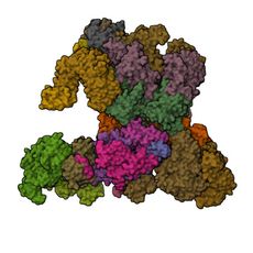
- PDB
- 7PMK
- Type
- protein + DNA
- Length
- 9,198 residues
- Chains
- 22
- Method
- electron microscopy
- Authors
- Jenkyn-Bedford, M., Yeeles, J.T.P., Deegan, T.D.
- Deposited
- 09/02/21
- Collection
- MolNFT v2 / PDB Bank
- Contract
- 0xd58B01f6C18086e5202cdC5D7Ad3E41790360102
- Owner
- 0xF4e1AA6f26B7DA48EeAD9B24a97927303eeA07b8
- Resolution
- 3.2
- Source
- Saccharomyces cerevisiae; SYNTHETIC CONSTRUCT
- Molecule
- DNA replication licensing factor MCM2; DNA replication licensing factor MCM3; DNA…
- EC
- 3.6.4.12, 2.7.7.7, 3.1.11.-
- UniProt
- A0A6A5Q1S9, P24279, P30665, A0A6A5PUY8, P53091, A0A6A5Q4N0, A0A6A5Q203, A0A6A5PX40, A0A6L1BWJ4, Q03406, Q08032, A0A6A5Q5Y5, A0A6A5Q435, Q08496, P21951, P24482, P53840, Q04659
- Ligands
- ANP, ZN, MG
- Pockets
- 20 · best druggability 0.87
Structure
electron microscopy · resolution 3.2 · 22 distinct chains · 42% helix, 20% strand (DSSP).
| Chain | Molecule | Type | Residues | Secondary structure | UniProt |
|---|---|---|---|---|---|
| 2 | DNA replication licensing factor MCM2 (EC 3.6.4.12) | protein | 655 | 43% helix, 22% strand | A0A6A5Q1S9 |
| 3 | DNA replication licensing factor MCM3 (EC 3.6.4.12) | protein | 607 | 38% helix, 24% strand | P24279 |
| 4 | DNA replication licensing factor MCM4 (EC 3.6.4.12) | protein | 597 | 39% helix, 24% strand | P30665 |
| 5 | DNA helicase (EC 3.6.4.12) | protein | 673 | 42% helix, 23% strand | A0A6A5PUY8 |
| 6 | DNA replication licensing factor MCM6 (EC 3.6.4.12) | protein | 637 | 35% helix, 24% strand | P53091 |
| 7 | DNA replication licensing factor MCM7 (EC 3.6.4.12) | protein | 629 | 40% helix, 21% strand | A0A6A5Q4N0 |
| A | DNA replication complex GINS protein PSF1 | protein | 197 | 58% helix, 11% strand | A0A6A5Q203 |
| B | DNA replication complex GINS protein PSF2 | protein | 192 | 49% helix, 8% strand | A0A6A5PX40 |
| C | DNA replication complex GINS protein PSF3 | protein | 171 | 58% helix, 9% strand | A0A6L1BWJ4 |
| D | DNA replication complex GINS protein SLD5 | protein | 243 | 53% helix, 11% strand | Q03406 |
| E | Cell division control protein 45,Cell division control protein 45 | protein | 564 | 50% helix, 15% strand | Q08032 |
| F | DNA polymerase alpha-binding protein | protein | 424 | 26% helix, 32% strand | A0A6A5Q5Y5 |
| G | DNA polymerase alpha-binding protein | protein | 422 | 27% helix, 34% strand | A0A6A5Q5Y5 |
| H | DNA polymerase alpha-binding protein | protein | 425 | 28% helix, 33% strand | A0A6A5Q5Y5 |
| I | Leading strand template DNA | dna | 36 | — | — |
| J | Lagging strand template DNA | dna | 33 | — | — |
| K | E3 ubiquitin ligase complex SCF subunit | protein | 137 | 50% helix, 8% strand | A0A6A5Q435 |
| L | Protein DIA2 | protein | 475 | 33% helix, 19% strand | Q08496 |
| Q | DNA polymerase epsilon catalytic subunit A (EC 2.7.7.7, 3.1.11.-) | protein | 766 | 43% helix, 19% strand | P21951 |
| R | DNA polymerase epsilon subunit B | protein | 552 | 34% helix, 22% strand | P24482 |
| X | Topoisomerase 1-associated factor 1 | protein | 665 | 68% helix, 3% strand | P53840 |
| Y | Chromosome segregation in meiosis protein 3 | protein | 98 | 64% helix, 0% strand | Q04659 |
Ligands and binding sites
- ANP · Phosphoaminophosphonic acid-adenylate ester · ligand · 3 copies · chains 2, 6, 3, 5
- ZN · Zinc ion · metal · 7 copies · chains 2, 4, 5, 6, 7, Q
- MG · Magnesium ion · metal · 3 copies · chains 2, 6, 3, 5
Binding sites are the residues within 4.5 Å of a ligand's heavy atoms.
Pockets
20 pockets found on a 1 Å grid, 20 with a druggability of 0.5 or more.
| Volume (ų) | Druggability | Buriedness | Lining residues | Inside | |
|---|---|---|---|---|---|
| 1 | 1065 | 0.87 | 0.88 | 144 | empty |
| 2 | 828 | 0.86 | 0.89 | 101 | empty |
| 3 | 694 | 0.86 | 0.90 | 98 | ANP, MG |
| 4 | 1105 | 0.85 | 0.86 | 110 | empty |
| 5 | 880 | 0.84 | 0.86 | 143 | empty |
Interfaces
70 interfaces between chains.
- 3–5 · 4860 Ų buried · 62 hydrogen bonds · protein-protein
- 2–6 · 4434 Ų buried · 56 hydrogen bonds · protein-protein
- 4–7 · 3856 Ų buried · 41 hydrogen bonds · protein-protein
- 4–6 · 3727 Ų buried · 40 hydrogen bonds · protein-protein
- 2–5 · 3501 Ų buried · 40 hydrogen bonds · protein-protein
Compared structures
- Q08032 · AlphaFold monomer V2.0 prediction for cell division control protein 45 (Q08032) · a predicted model of the same protein · chain E against A, TM-score 0.99
- P21951 · AlphaFold monomer V2.0 prediction for DNA polymerase epsilon catalytic subunit a (P21951) · a predicted model of the same protein · chain Q against A, TM-score 0.97
- Q08496 · AlphaFold monomer V2.0 prediction for protein DIA2 (Q08496) · a predicted model of the same protein · chain L against A, TM-score 0.96
- P24279 · AlphaFold monomer V2.0 prediction for DNA replication licensing factor MCM3 (P24279) · a predicted model of the same protein · chain 3 against A, TM-score 0.96
- P30665 · AlphaFold monomer V2.0 prediction for DNA replication licensing factor MCM4 (P30665) · a predicted model of the same protein · chain 4 against A, TM-score 0.92
- P53840 · AlphaFold monomer V2.0 prediction for topoisomerase 1-associated factor 1 (P53840) · a predicted model of the same protein · chain X against A, TM-score 0.91
- Q03406 · AlphaFold monomer V2.0 prediction for DNA replication complex gins protein SLD5 (Q03406) · a predicted model of the same protein · chain D against A, TM-score 0.91
- Q04659 · AlphaFold monomer V2.0 prediction for chromosome segregation in meiosis protein 3 (Q04659) · a predicted model of the same protein · chain Y against A, TM-score 0.86
- P24482 · AlphaFold monomer V2.0 prediction for DNA polymerase epsilon subunit B (P24482) · a predicted model of the same protein · chain R against A, TM-score 0.85
- P53091 · AlphaFold monomer V2.0 prediction for DNA replication licensing factor MCM6 (P53091) · a predicted model of the same protein · chain 6 against A, TM-score 0.84
Analyse
- Open in the research workbench: secondary structure, accessibility, pockets, interfaces, conservation, variants, modifications, peptides and crosslinks on the 3D structure
- Every structure of UniProt A0A6A5Q1S9
- Every structure of UniProt P24279
- Every structure of UniProt P30665
- Every structure of UniProt A0A6A5PUY8
- Every structure of UniProt P53091
- Every structure of UniProt A0A6A5Q4N0
- Every structure of UniProt A0A6A5Q203
- Every structure of UniProt A0A6A5PX40
- Every structure of UniProt A0A6L1BWJ4
- Every structure of UniProt Q03406
- Every structure of UniProt Q08032
- Every structure of UniProt A0A6A5Q5Y5
- Every structure of UniProt A0A6A5Q435
- Every structure of UniProt Q08496
- Every structure of UniProt P21951
- Every structure of UniProt P24482
- Every structure of UniProt P53840
- Every structure of UniProt Q04659
- Elsewhere: RCSB PDB 7PMK · PDBe 7PMK · UniProt A0A6A5Q1S9 · UniProt P24279 · UniProt P30665 · UniProt A0A6A5PUY8 · UniProt P53091 · UniProt A0A6A5Q4N0 · UniProt A0A6A5Q203 · UniProt A0A6A5PX40 · UniProt A0A6L1BWJ4 · UniProt Q03406 · UniProt Q08032 · UniProt A0A6A5Q5Y5 · UniProt A0A6A5Q435 · UniProt Q08496 · UniProt P21951 · UniProt P24482 · UniProt P53840 · UniProt Q04659
GL1X is a live explorer of the GenesisL1 blockchain. Turn on JavaScript for search, charts and interactive views.