MolNFT v2 · #159827 · protein
6O2T: Acetylated Microtubules
6O2T · Tubulin alpha-1B chain: “Acetylated Microtubules”. Electron microscopy at 4.1, 3 ligands (GTP, GDP, MG), 20 pockets (best druggability 0.86). MolNFT v2 #159827 on GenesisL1: 3D structure, binding sites and pockets in GL1X.
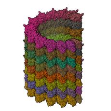
- PDB
- 6O2T
- Type
- protein
- Length
- 45,032 residues
- Chains
- 104
- Method
- electron microscopy
- Authors
- Eshun-Wilson, L., Zhang, R., Portran, D., Nachury, M.V., Toso, D., Lohr, T., Vendruscolo, M., Bonomi, M., Fraser, J.S., Nogales, E.
- Deposited
- 02/24/19
- Collection
- MolNFT v2 / PDB Bank
- Contract
- 0xd58B01f6C18086e5202cdC5D7Ad3E41790360102
- Owner
- 0xF4e1AA6f26B7DA48EeAD9B24a97927303eeA07b8
- Resolution
- 4.1
- Source
- Sus scrofa
- Molecule
- Tubulin alpha-1B chain; Tubulin beta chain
- UniProt
- Q2XVP4, P02554
- Ligands
- GTP, GDP, MG
- Pockets
- 20 · best druggability 0.86
Structure
electron microscopy · resolution 4.1 · 2 distinct chains · 44% helix, 17% strand (DSSP).
| Chain | Molecule | Type | Residues | Secondary structure | UniProt |
|---|---|---|---|---|---|
| 1A, 1B, 1C, 1D, 1E, 1F, 1G, 1I, 1J, 1K, 1L, 1M, 1N, 2A, 2B, 2C, 2D, 2E, 2F, 2G, 2I, 2J, 2K, 2L, 2M, 2N, 3A, 3B, 3C, 3D, 3E, 3F, 3G, 3I, 3J, 3K, 3L, 3M, 3N, 4A, 4B, 4C, 4D, 4E, 4F, 4G, 4I, 4J, 4K, 4L, 4M, 4N | Tubulin alpha-1B chain | protein | 437 | 43% helix, 17% strand | Q2XVP4 |
| 1H, 1O, 1P, 1Q, 1R, 1S, 1T, 1U, 1V, 1W, 1X, 1Y, 1Z, 2H, 2O, 2P, 2Q, 2R, 2S, 2T, 2U, 2V, 2W, 2X, 2Y, 2Z, 3H, 3O, 3P, 3Q, 3R, 3S, 3T, 3U, 3V, 3W, 3X, 3Y, 3Z, 4H, 4O, 4P, 4Q, 4R, 4S, 4T, 4U, 4V, 4W, 4X, 4Y, 4Z | Tubulin beta chain | protein | 429 | 44% helix, 18% strand | P02554 |
Ligands and binding sites
- GTP · Guanosine-5'-triphosphate · ligand · 52 copies · chains 1A, 1H, 1B, 1O, 1C, 1P, 1D, 1Q, 1E, 1R, 1F, 1S, 1G, 1T, 1I, 1U, 1J, 1V, 1K, 1W, 1L, 1X, 1M, 1Y, 1N, 1Z, 2A, 2H, 2B, 2O, 2C, 2P, 2D, 2Q, 2E, 2R, 2F, 2S, 2G, 2T, 2I, 2U, 2J, 2V, 2K, 2W, 2L, 2X, 2M, 2Y, 2N, 2Z, 3A, 3H, 3B, 3O, 3C, 3P, 3D, 3Q, 3E, 3R, 3F, 3S, 3G, 3T, 3I, 3U, 3J, 3V, 3K, 3W, 3L, 3X, 3M, 3Y, 3N, 3Z, 4A, 4H, 4B, 4O, 4C, 4P, 4D, 4Q, 4E, 4R, 4F, 4S, 4G, 4T, 4I, 4U, 4J, 4V, 4K, 4W, 4L, 4X, 4M, 4Y, 4N, 4Z
- GDP · Guanosine-5'-diphosphate · ligand · 52 copies · chains 1H, 2A, 1O, 2B, 1P, 2C, 1Q, 2D, 1R, 2E, 1S, 2F, 1T, 2G, 1U, 2I, 1V, 2J, 1W, 2K, 1X, 2L, 1Y, 2M, 1Z, 2N, 2H, 3A, 2O, 3B, 2P, 3C, 2Q, 3D, 2R, 3E, 2S, 3F, 2T, 3G, 2U, 3I, 2V, 3J, 2W, 3K, 2X, 3L, 2Y, 3M, 2Z, 3N, 3H, 3O, 3P, 3Q, 3R, 3S, 3T, 3U, 3V, 3W, 3X, 3Y, 3Z, 1A, 4H, 1B, 4O, 1C, 4P, 1D, 4Q, 1E, 4R, 1F, 4S, 1G, 4T, 1I, 4U, 1J, 4V, 1K, 4W, 1L, 4X, 1M, 4Y, 1N, 4Z
- MG · Magnesium ion · metal · 52 copies · chains 1A, 1B, 1C, 1D, 1E, 1F, 1G, 1I, 1J, 1K, 1L, 1M, 1N, 2A, 2B, 2C, 2D, 2E, 2F, 2G, 2I, 2J, 2K, 2L, 2M, 2N, 3A, 3B, 3C, 3D, 3E, 3F, 3G, 3I, 3J, 3K, 3L, 3M, 3N, 4A, 4B, 4C, 4D, 4E, 4F, 4G, 4I, 4J, 4K, 4L, 4M, 4N
Binding sites are the residues within 4.5 Å of a ligand's heavy atoms.
Pockets
20 pockets found on a 1 Å grid, 20 with a druggability of 0.5 or more.
| Volume (ų) | Druggability | Buriedness | Lining residues | Inside | |
|---|---|---|---|---|---|
| 1 | 254 | 0.86 | 0.98 | 48 | GDP |
| 2 | 258 | 0.85 | 0.97 | 46 | GDP |
| 3 | 242 | 0.85 | 0.97 | 44 | GDP |
| 4 | 221 | 0.85 | 0.98 | 44 | GDP |
| 5 | 246 | 0.84 | 0.97 | 48 | GDP |
Interfaces
192 interfaces between chains.
- 1D–4Q · 2218 Ų buried · 71 hydrogen bonds · protein-protein
- 1C–4P · 2213 Ų buried · 68 hydrogen bonds · protein-protein
- 1E–4R · 2203 Ų buried · 73 hydrogen bonds · protein-protein
- 1B–4O · 2191 Ų buried · 64 hydrogen bonds · protein-protein
- 1F–4S · 2181 Ų buried · 69 hydrogen bonds · protein-protein
Compared structures
- Q2XVP4 · AlphaFold monomer V2.0 prediction for tubulin alpha-1B chain (Q2XVP4) · a predicted model of the same protein · chain 1A against A, TM-score 0.99
- P02554 · AlphaFold monomer V2.0 prediction for tubulin beta chain (P02554) · a predicted model of the same protein · chain 1H against A, TM-score 0.98
Analyse
- Open in the research workbench: secondary structure, accessibility, pockets, interfaces, conservation, variants, modifications, peptides and crosslinks on the 3D structure
- Every structure of UniProt Q2XVP4
- Every structure of UniProt P02554
- Elsewhere: RCSB PDB 6O2T · PDBe 6O2T · UniProt Q2XVP4 · UniProt P02554
GL1X is a live explorer of the GenesisL1 blockchain. Turn on JavaScript for search, charts and interactive views.