MolNFT v2 · #196145 · protein + DNA
7U1P: RFC:PCNA bound to DNA with a ssDNA gap of five nucleotides
7U1P · Replication factor C subunit 1: “RFC:PCNA bound to DNA with a ssDNA gap of five nucleotides”. Electron microscopy at 3.0, 3 ligands (AGS, ADP, MG), 20 pockets (best druggability 0.88). MolNFT v2 #196145 on GenesisL1: 3D structure…
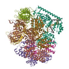
- PDB
- 7U1P
- Type
- protein + DNA
- Length
- 2,769 residues
- Chains
- 11
- Method
- electron microscopy
- Authors
- Liu, X., Gaubitz, C., Pajak, J., Kelch, B.A.
- Deposited
- 02/21/22
- Collection
- MolNFT v2 / PDB Bank
- Contract
- 0xd58B01f6C18086e5202cdC5D7Ad3E41790360102
- Owner
- 0xF4e1AA6f26B7DA48EeAD9B24a97927303eeA07b8
- Resolution
- 3.0
- Source
- Saccharomyces cerevisiae; SYNTHETIC CONSTRUCT
- Molecule
- Replication factor C subunit 1; Replication factor C subunit 4; Replication factor C…
- UniProt
- P38630, P40339, P38629, P40348, P38251, P15873
- Ligands
- AGS, ADP, MG
- Pockets
- 20 · best druggability 0.88
Structure
electron microscopy · resolution 3.0 · 11 distinct chains · 46% helix, 19% strand (DSSP).
| Chain | Molecule | Type | Residues | Secondary structure | UniProt |
|---|---|---|---|---|---|
| A | Replication factor C subunit 1 | protein | 617 | 56% helix, 8% strand | P38630 |
| B | Replication factor C subunit 4 | protein | 319 | 61% helix, 8% strand | P40339 |
| C | Replication factor C subunit 3 | protein | 328 | 57% helix, 9% strand | P38629 |
| D | Replication factor C subunit 2 | protein | 340 | 61% helix, 9% strand | P40348 |
| E | Replication factor C subunit 5 | protein | 338 | 53% helix, 13% strand | P38251 |
| F | Proliferating cell nuclear antigen | protein | 255 | 18% helix, 43% strand | P15873 |
| G | Proliferating cell nuclear antigen | protein | 257 | 18% helix, 44% strand | P15873 |
| H | Proliferating cell nuclear antigen | protein | 253 | 17% helix, 42% strand | P15873 |
| I | DNA - Primer | dna | 18 | — | — |
| J | DNA - Template | dna | 34 | — | — |
| K | DNA - non primer | dna | 10 | — | — |
Ligands and binding sites
- AGS · Phosphothiophosphoric acid-adenylate ester · ligand · 3 copies · chains A, B, C, D, E
- ADP · Adenosine-5'-diphosphate · ligand · 2 copies · chains B, C, A, E
- MG · Magnesium ion · metal · 4 copies · chains A, B, C, D, E
Binding sites are the residues within 4.5 Å of a ligand's heavy atoms.
Pockets
20 pockets found on a 1 Å grid, 20 with a druggability of 0.5 or more.
| Volume (ų) | Druggability | Buriedness | Lining residues | Inside | |
|---|---|---|---|---|---|
| 1 | 1041 | 0.88 | 0.89 | 131 | empty |
| 2 | 806 | 0.84 | 0.87 | 102 | empty |
| 3 | 201 | 0.81 | 0.95 | 32 | ADP, MG |
| 4 | 700 | 0.81 | 0.85 | 54 | empty |
| 5 | 183 | 0.80 | 0.95 | 33 | AGS, MG |
Interfaces
34 interfaces between chains.
- A–E · 2984 Ų buried · 31 hydrogen bonds · protein-protein
- D–E · 2932 Ų buried · 43 hydrogen bonds · protein-protein
- B–C · 2731 Ų buried · 37 hydrogen bonds · protein-protein
- C–D · 2563 Ų buried · 30 hydrogen bonds · protein-protein
- A–B · 2442 Ų buried · 28 hydrogen bonds · protein-protein
Compared structures
- 7U1P · RFC:PCNA bound to DNA with a ssdna gap of five nucleotides · the same entry in another collection · chain K against K, TM-score 1.00
- P15873 · AlphaFold monomer V2.0 prediction for proliferating cell nuclear antigen (P15873) · a predicted model of the same protein · chain F against A, TM-score 0.98
Analyse
- Open in the research workbench: secondary structure, accessibility, pockets, interfaces, conservation, variants, modifications, peptides and crosslinks on the 3D structure
- Every structure of UniProt P38630
- Every structure of UniProt P40339
- Every structure of UniProt P38629
- Every structure of UniProt P40348
- Every structure of UniProt P38251
- Every structure of UniProt P15873
- Elsewhere: RCSB PDB 7U1P · PDBe 7U1P · UniProt P38630 · UniProt P40339 · UniProt P38629 · UniProt P40348 · UniProt P38251 · UniProt P15873
Other records of 7U1P
- 7U1P-2 · MolNFT v1 / PDB · RFC:PCNA bound to DNA with a ssdna gap of five nucleotides
GL1X is a live explorer of the GenesisL1 blockchain. Turn on JavaScript for search, charts and interactive views.