MolNFT v2 · #92557 · protein
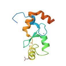
4GB0: Crystal Structure of the RING domain of RNF168
4GB0 · E3 ubiquitin-protein ligase RNF168: “Crystal Structure of the RING domain of RNF168”. X-ray diffraction at 2.6, 1 ligand (ZN). MolNFT v2 #92557 on GenesisL1: 3D structure, binding sites and pockets in GL1X.
- PDB
- 4GB0
- Type
- protein
- Length
- 91 residues
- Chains
- 1
- Method
- x-ray diffraction
- Authors
- Zhang, X.Q., Wang, C.L., Zang, J.Y.
- Deposited
- 07/26/12
- Collection
- MolNFT v2 / PDB Bank
- Contract
- 0xd58B01f6C18086e5202cdC5D7Ad3E41790360102
- Owner
- 0xF4e1AA6f26B7DA48EeAD9B24a97927303eeA07b8
- Resolution
- 2.6
- Source
- Homo sapiens
- Molecule
- E3 ubiquitin-protein ligase RNF168
- EC
- 6.3.2.-
- UniProt
- Q8IYW5
- Ligands
- ZN
Structure
x-ray diffraction · resolution 2.6 · 1 distinct chain · 46% helix, 11% strand (DSSP).
| Chain | Molecule | Type | Residues | Secondary structure | UniProt |
|---|---|---|---|---|---|
| A | E3 ubiquitin-protein ligase RNF168 (EC 6.3.2.-) | protein | 91 | 46% helix, 11% strand | Q8IYW5 |
Ligands and binding sites
Binding sites are the residues within 4.5 Å of a ligand's heavy atoms.
Compared structures
Analyse
- Open in the research workbench: secondary structure, accessibility, pockets, interfaces, conservation, variants, modifications, peptides and crosslinks on the 3D structure
- Every structure of UniProt Q8IYW5
- Elsewhere: RCSB PDB 4GB0 · PDBe 4GB0 · UniProt Q8IYW5
Other records of 4GB0
- 4GB0-2 · MolNFT v1 / PDB · Crystal structure of the ring domain of RNF168
GL1X is a live explorer of the GenesisL1 blockchain. Turn on JavaScript for search, charts and interactive views.