MolNFT v1 · #7258 · protein
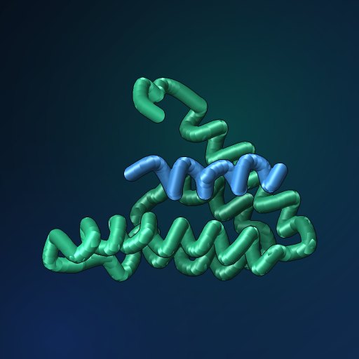
1G1E: NMR structure of the human MAD1 transrepression domain sid in complex with mammalian SIN3A PAH2 domain
1G1E · MAD1 protein: “NMR structure of the human MAD1 transrepression domain sid in complex with mammalian SIN3A PAH2 domain”. Solution nmr. MolNFT v1 #7258 on GenesisL1: 3D structure, binding sites and pockets in GL1X.
- PDB
- 1G1E
- Type
- protein
- Length
- 105 residues
- Chains
- 2
- Method
- solution nmr
- Deposited
- 11-OCT-00
- Collection
- MolNFT v1 / PDB
- Contract
- 0xDE3723766Bc32dcACD03C17BaA400A7B36837Eba
- Owner
- 0x3396CDB397284571d12822B4ae1189a8E22AAAAA
- Molecule
- MAD1 protein; SIN3A
- UniProt
- Q60520
Structure
solution nmr · 2 distinct chains · 68% helix, 0% strand (DSSP).
| Chain | Molecule | Type | Residues | Secondary structure | UniProt |
|---|---|---|---|---|---|
| A | MAD1 protein | protein | 16 | 81% helix, 0% strand | — |
| B | SIN3A | protein | 89 | 65% helix, 0% strand | Q60520 |
Interfaces
1 interface between chains.
- A–B · 804 Ų buried · 2 hydrogen bonds · protein-protein
Compared structures
- 1G1E · NMR structure of the human MAD1 transrepression domain sid in complex with mammalian… · the same entry in another collection · chain A against A, TM-score 1.00
- Q60520 · AlphaFold monomer V2.0 prediction for paired amphipathic helix protein SIN3A (Q60520) · a predicted model of the same protein · chain B against A, TM-score 0.79
Analyse
- Open in the research workbench: secondary structure, accessibility, pockets, interfaces, conservation, variants, modifications, peptides and crosslinks on the 3D structure
- Every structure of UniProt Q60520
- Elsewhere: RCSB PDB 1G1E · PDBe 1G1E · UniProt Q60520
Other records of 1G1E
- 1G1E · MolNFT v2 / PDB Bank · NMR structure of the human MAD1 transrepression domain sid in complex with mammalian…
GL1X is a live explorer of the GenesisL1 blockchain. Turn on JavaScript for search, charts and interactive views.